Histology & Histopathology

An Accompanying Resource to the Laboratory Manual

This website serves as a histology and histopathology resource for students studying for lab tests, midterms, and exams.

Purpose

Histology refers to the study of normal cells whereas histopathology explores the transformation of normal structures in response to diseases such as cancer and inflammation.

This resource explores the interplay between structures of different organs and tissues and their function. It is a compilation of notes from the lectures, tutorial slides, lab manual, and Wheater’s Functional Histology, 6e.

Sample Panoramic Images

Sample chapter module

Digestive System

Stomach

1. Mucosa

Epithelium | simple columnar epithelium

Cellular component | surface lining cells, parietal cells, regenerative cells, mucous neck cells, chief cells, enteroendocrine cells, NO goblet cells

Lamina Propria | loose connective tissue, gastric glands (simple tubular)

Muscularis Mucosae | IC/OL SMC layer & third circular SMC layer

2. Submucosa

Connective Tissue | dense irregular collagenous connective tissue

3. Muscularis Externa

Inner oblique SMC

Middle circular SMC

OL SMC

Stomach

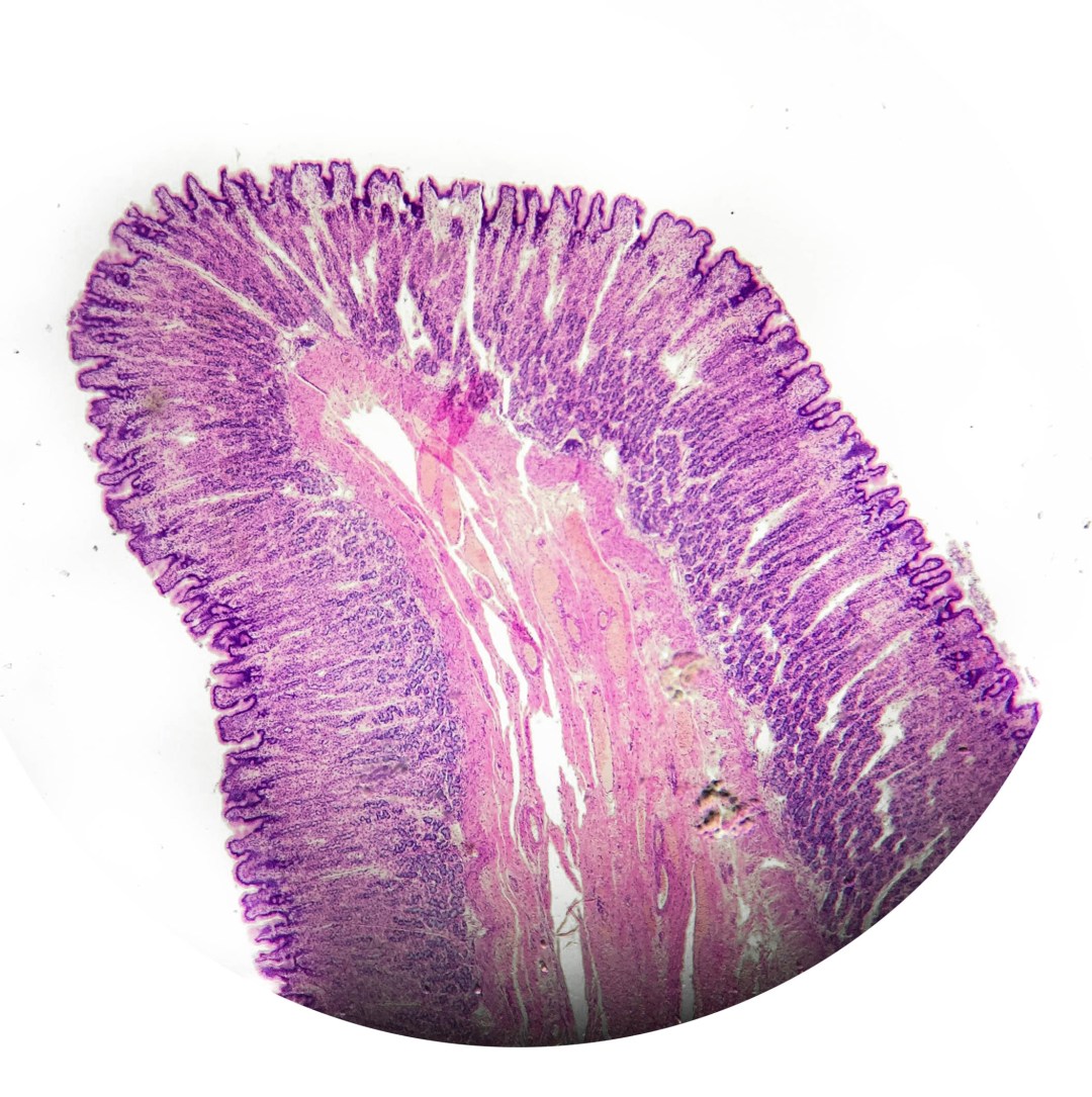
A view of the mucosa in a folded state showing the gastric glands extending from the lumen into the muscularis mucosae.

Stomach

Another look at the gastric glands which are straight tubular glands synthesizing gastric juice. The next photo shows a closer look at the epithelium layer.

Stomach

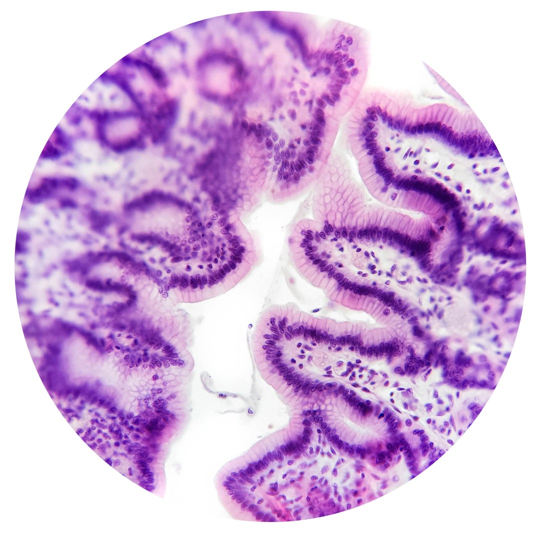
A closer view of the simple columnar epithelium lining the mucosa. Note the nuclei are all close to the basement membrane.

Stomach

This shows the thickness of the gastric body in the mucosa and a small part of the submucosa. The gastric pits are evident. Look at the next photo for a closer view of the mucous neck cells and parietal cells. Although very small, the chief cells can be found near the base of the gastric glands, which extend down into the muscularis mucosae. Notice how the normal gastric mucosa has no lymphoid cells.

Stomach

Here, you can see the parietal cells (magenta colour with central nuclei) lining the lower part of the gastric pits. Again, the simple columnar epithelium is evident near the top. You may also spot some mucous neck cells which are more pale in appearance. Not seen here, are the chief cells which are located close to the bottoms of the gastric pits.

SAMPLE DIAGRAMS

DIGESTIVE system

circulatory system

authors & contributors

Dr. Ron Wilson Jr.

Dr. Ron Wilson Jr.

Professor

Ingrid Barany

Ingrid Barany

Website Designer & Author

Max Shcherbina

Max Shcherbina

Website Designer & Author

SEND us feedback & suggestions!

6 + 4 =