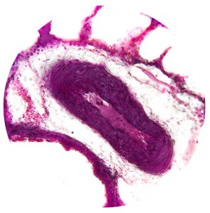
Tissues & Organs
Tissues & Organs
General Histology of a Vessel
1. Tunica Intima
Endothelial layer
Basement membrane
Subendothelial connective tissue
Internal elastic lamina
2. Tunica Media
Layers of smooth muscle cells, elastic fibres, type III collagen, and proteoglycans
External lamina
Vasa vasorum & nervi vasorum
3. Tunica Adventitia
Loose connective tissue
Fibroblasts, type I collagen, and elastic fibres
Arteries
Elastic Arteries | Conducting
1. Tunica Intima
Endothelial layer | contains Weibel-Palade bodies
Subendothelial Connective Tissue | narrow
Internal Elastic Lamina | thin/incomplete
2. Tunica Media
Layers of elastic fibres | 40-70
Layers of Circular Smooth Muscle Cells | longer than normal
External Lamina | thin
Vasa Vasorum | may be present
3. Tunica Adventitia
Loose Connective Tissue | thin
Fibroblasts, type I collagen, and elastic fibres
Vasa Vasorum, nervi vasorum, lymphatic vessels
Muscular Arteries | Distributing
1. Tunica Intima
Endothelial layer
Subendothelial Connective Tissue | thinner than elastic arteries
Internal Elastic Lamina | distinct, may be bifid
2. Tunica Media
Layers of Elastic Fibres | 4 (small arteries) – 40 (larger arteries)
Layers of circular smooth muscle cells, elastic fibres, and type III collagen
External Lamina | present
Vasa Vasorum | may be present
3. Tunica Adventitia
Fibroblasts, type I collagen, and elastic fibres
Vasa vasorum, nervi vasorum

Artery & Vein

Blood Vessels, Trachea & Esophagus

Atherosclerosis
Arterioles
1. Tunica Intima
Endothelial Layer | thin
Subendothelial Connective Tissue | type III collagen and elastic fibres
Internal Elastic Lamina | thin and fenestrated (larger arterioles), absent (small arterioles)
2. Tunica Media
Layers of Elastic Fibres | 1 (small arterioles) – 3 (larger arterioles)
Layers of circular smooth muscle cells, elastic fibres, and type III collagen
External Lamina | absent
3. Tunica Adventitia
Fibroblastic Connective Tissue | few fibroblasts
Veins
Large Veins
Lumen
Usually collapsed due to thin walls and less elastic fibres in comparison to arteries
1. Tunica Intima
Endothelial Layer
Subendothelial Connective Tissue | thick, contains reticular fibres, elastic fibres, and fibroblasts
Internal Elastic Lamina | present
2. Tunica Media
Usually absent
3. Tunica Adventitia
Loose Connective Tissue | Type I collagen, and elastic fibres
Muscle Layer | longitudinal smooth muscle cells (inferior vena cava), cardiac muscle (pulmonary veins and Vena Cava)
Vasa Vasorum
Medium Veins
1. Tunica Intima
Endothelial Layer
Subendothelial Connective Tissue | contains reticular fibres, elastic fibres, and fibroblasts
Internal Elastic Lamina | absent
2. Tunica Media
Muscle Layer | smooth muscle cells interwoven with collagen and fibroblasts
3. Tunica Adventitia
Muscle Layer | longitudinal collagen and elastic fibres, with few smooth muscle cells
Venules & Small Veins
1. Tunica Intima
Endothelial Layer | thin
Subendothelial Connective Tissue | reticular fibres and pericytes
Capillaries
Epithelium | single layer of endothelial cells surrounded by pericytes


